“Fear the robots.” Those three words have long been the caveat and cornerstone of classic science fiction.
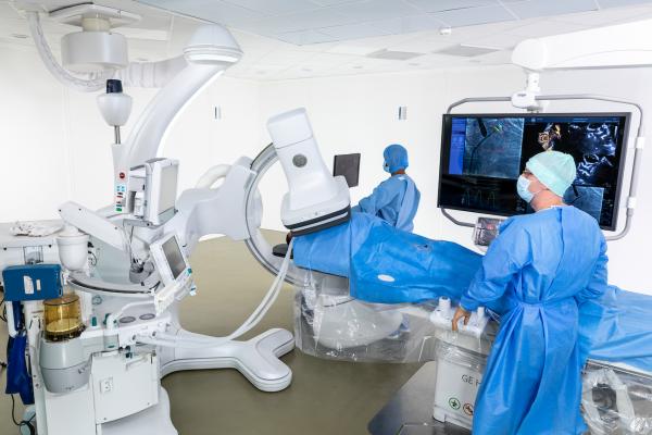
But in the angiography suite, robotics has received a decidedly warmer reception since debuting more than a decade ago. Simply defined, robotic angiography involves any medical imaging device that leverages some form of robotics — a mechanical arm, for example, or a small, motorized vehicle — to aid with procedures that otherwise would fall to a human in the angiography suite.
Interest in robotic angiography is rising, thanks to accelerated growth in minimally invasive procedures performed in the angiography suite. In interventional radiology (IR), these procedures are primarily tumor embolization and ablation. In the cardiovascular space, they include structural heart procedures for artificial valve replacement of the aortic and tricuspid valves as well as complex endovascular procedures such as abdominal aortic aneurysms. During these IR and cardiovascular procedures, robotic angiography helps improve physical access to patients, facilitate advanced imaging, and control infection.
Regarding patient access, robotic angiography allows for unmatched flexibility with head-to-toe coverage. Radial artery access is a rising trend within the interventional cardiology community, and robotic angiography allows clinicians to obtain images without sacrificing patient safety by rotating the patient table laterally to allow for easy access to the patient’s wrists without compromising workflow. During a mitral valve procedure, a robotic angiography system permits the interventional cardiology team to work with ultrasound and anesthesia teams to provide critical information to guide the replacement valve or device, without interference from the movements of the angiography system. And in an endovascular abdominal aortic aneurysm, the wide C-arm of some robotic angiography units enables vascular surgeons to safely access larger patients while working with devices and instruments.
On the advanced imaging front, robotic angiography possesses unique capabilities in rotational angiography and fusion imaging. Allowing spins from multiple positions around the patient table, rotational angiography (aka 3D cone beam computed tomography) is increasingly used for real-time confirmation in IR oncology procedures such as liver tumor embolization. In structural heart procedures, robotic angiography systems provide new levels of integration and workflow, enabling simultaneous fusion of CT, ultrasound, and angiography. And in vascular procedures, CT fusion imaging enables physicians to treat patients with less radiation exposure and more efficiently (i.e., for complex fenestrated stent grafts).
In the infection control arena, some robotic angiography units allow for an operating room (OR) type of environment by avoiding contact with the angiography suite’s ceiling and floor. This OR-like environment permits necessary laminar air flow over the patient while limiting contact with bodily fluids, cabling, and medical equipment.
Expect use of robotic angiography systems to expand in the coming years, in part because more surgical procedures will become minimally invasive. In the IR space, for example, more embolization and ablation procedures will be used to treat new conditions. These procedures will potentially include bariatric embolization, prostate artery embolization, and osteoarthritis embolization (aka geniculate embolization), or the embolization of blood vessels connected to nerves in the knees to prevent joint pain. In the structural heart realm, repair of mitral and tricuspid valvular disease eventually will be as commonplace as transcatheter aortic valve repair (TAVR), bringing relief to an underserved patient population that currently undergoes highly complex procedures at higher-acuity institutions.